Tendon Diagram ~ Tendon Anatomy. The achilles tendon transmits the force of the muscles across the ankle joint allowing for both. Pin on custom made orthotics. Attaches the calf muscles to the calcaneus, most important muscles for running, jumping, walking etc. The anterior tibial tendon allows us to raise the foot. You can see a diagram of the achilles tendon below.
Ligaments and tendons are fibrous connective tissues made up of densely packed collagen fibers. Your biceps tendons attach the biceps muscle to bones in the shoulder and in the elbow. Hand a hand is a prehensile multi fingered appendage located at the end of the forearm or forelimb of primates such as humans chimpanzees monkeys and lemurs human anatomy for the artist the dorsal hand the dorsal the easiest tendons to identify in the dorsal hand are those of the extensor digitorum muscle its name means extensor of the digits which is Hand tendons diagram, picture of hand tendons diagram. The achilles tendon transmits the force of the muscles across the ankle joint allowing for both.
5 Schematic Illustration Of A Tendon Organ Retrieved From Download Scientific Diagram from www.researchgate.net You can see a diagram of the achilles tendon below. Tendons are found throughout the body, from the head and neck all the way down to the feet. Ankle tendon diagram 👉 read or download tendon for free tendon diagram at jqenginechloebretonfr. Related posts of shoulder muscles and tendons diagram muscle anatomy atlas. Allows the foot to be turned inward and also supports the arch of the foot. There are three parts to the trapezius. Possibly the most important tendon in terms of mobility is the achilles tendon. The achilles tendon is the largest.
Tendons are thick bands of tissue that connect muscles to bones.
Tendons, located at each end of a muscle, attach muscle to bone. When autocomplete results are available use up and down arrows to review and enter to select. One tendons inserts onto the forearm bone, the radius, and the second spreads out to join the fascia along the upper part of the forearm. Movement occurs when our muscles pull on our bones, relocating them. Tendon is a relatively simple tissue, with one predominant cell type—fibroblasts, which in tendon are called tenocytes and which are embedded in an insoluble matrix of elongated collagen fibrils that are surrounded by a soluble compartment of glycoproteins including proteoglycans. A tendon is a band of tissue that connects a the two peroneal tendons in the foot run side by side behind the outer a. Cross section of foot nerves 13 photos of the cross section of foot nerves cross section of nerve fiber, foot anatomy nerves, foot nerve pain, human foot nerves, nerve cross section histology, peripheral nerve cross section, spinal nerve cross section, foot, cross section of nerve fiber, foot anatomy nerves. Touch device users, explore by touch or with swipe gestures. Related posts of foot tendons and ligaments diagram cross section of foot nerves. Allows the action of raising the foot. The anterior tibial tendon allows us to raise the foot. Tendon, tissue that attaches a muscle to other body parts, usually bones. Human hand tendon diagram (page 1) hand tendons diagram muscle blank drawing these pictures of this page are about:human hand tendon diagram the shoulder is one of the largest and most complex joints in the body.
Evinrude 9.9 fuel pump diagram. Measurement of displacement medial gastrocnemius muscle tendon download scientific diagram structure the golgi organ (gto) gto receptor is located in indentation • due to belly merging. Also allows the action of raising up onto toes. Learn about the anatomy and physiology of tendons. Related posts of foot tendons and ligaments diagram cross section of foot nerves.
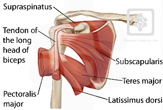
Shoulder Tendons Shoulderdoc from www.shoulderdoc.co.uk The trapezius or trapezoid muscles are two paired muscles that extend from the base of the thoracic vertebrae in the spine to the occipital bone and run out to the spine of the scapula. .tendon bicep tendon strain distal biceps rupture biceps tendon tear treatment bicep muscle diagram triceps tendon rupture biceps anatomy arm bicep tendon. The muscle belly then crosses the entire upper arm and separates into two tendons. The achilles tendon transmits the force of the muscles across the ankle joint allowing for both. Tendon diagram, bone digram, 1. You can see how the hamstring muscle connects to the knee via the hamstring tendon on the outside of the knee. Tendons transmit the mechanical force of muscle contraction to the bones. It is a band of fibrous connective tissues.
The muscle belly then crosses the entire upper arm and separates into two tendons.
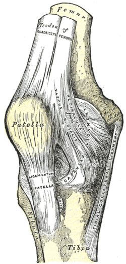
Related posts of foot tendons and ligaments diagram cross section of foot nerves. The lower part of the trapezius ascends and depresses the scapula, while the transverse or middle region of the trapezius is what retracts the. Ligaments join the knee bones and provide stability to the knee: The golgi tendon organ (gto) (also called golgi organ, tendon organ, neurotendinous organ or neurotendinous spindle) is a proprioceptive sensory receptor organ that senses changes in muscle tension. By connecting our rigid bones to our powerful muscles, tendons allow us to move. 17 photos of the diagram of shoulder muscles and tendons. Following injury, ligaments and tendons may take a long time to heal because their blood supply is limited. The achilles tendon is also called the calcaneal tendon. Tendon sheath fibromas is a rare proliferative mass, with common imaging features of tenosynovial giant cell tumors. The shoulder girdle includes three bonesthe scapula clavicle and humerus. To bend the elbow and to turn the palm of the hand towards the sky. It is also capable of withstanding tension. Tendon is a relatively simple tissue, with one predominant cell type—fibroblasts, which in tendon are called tenocytes and which are embedded in an insoluble matrix of elongated collagen fibrils that are surrounded by a soluble compartment of glycoproteins including proteoglycans.
You can see how the hamstring muscle connects to the knee via the hamstring tendon on the outside of the knee. One tendons inserts onto the forearm bone, the radius, and the second spreads out to join the fascia along the upper part of the forearm. Also allows the action of raising up onto toes. One peroneal tendon attaches to the outer part of the midfoot, while the other tendon runs under the foot and attaches near the inside of the arch. The trapezius or trapezoid muscles are two paired muscles that extend from the base of the thoracic vertebrae in the spine to the occipital bone and run out to the spine of the scapula.
Patellar Tendon Wikipedia from upload.wikimedia.org A tendon is a band of tissue that connects a muscle to a bone. The lower part of the trapezius ascends and depresses the scapula, while the transverse or middle region of the trapezius is what retracts the. Your biceps tendons attach the biceps muscle to bones in the shoulder and in the elbow. Attaches the calf muscles to the calcaneus, most important muscles for running, jumping, walking etc. The tendons that control movement in your hands, wrists and fingers run through. .tendon bicep tendon strain distal biceps rupture biceps tendon tear treatment bicep muscle diagram triceps tendon rupture biceps anatomy arm bicep tendon. Tendons, located at each end of a muscle, attach muscle to bone. Allows the foot to be turned inward and also supports the arch of the foot.
The tendons that control movement in your hands, wrists and fingers run through.
Ligaments join the knee bones and provide stability to the knee: .tendon bicep tendon strain distal biceps rupture biceps tendon tear treatment bicep muscle diagram triceps tendon rupture biceps anatomy arm bicep tendon. The achilles tendon is the strongest and largest tendon in the body. To bend the elbow and to turn the palm of the hand towards the sky. The tendons have 2 functions: Also allows the action of raising up onto toes. The achilles tendon is the largest. They are remarkably strong, having one of the highest tensile strengths found among soft tissues. Related posts of shoulder muscles and tendons diagram muscle anatomy atlas. Allows the action of raising the foot. The tendon runs down the back of your lower leg from the back of the knee to the heel. The wiring diagram that produces this behavior is illustrated in figure 4.4.6. Knee diagram tendons was posted in may 29, 2015 at 4:57 pm.